Home » Without Label » Abdominal Muscle Anatomy Male / 2 | Male Abdominal Muscle Anatomy Prints, Genuine 1922 ... - Huge collection, amazing choice, 100+ million high quality, affordable rf and rm images.
Abdominal Muscle Anatomy Male / 2 | Male Abdominal Muscle Anatomy Prints, Genuine 1922 ... - Huge collection, amazing choice, 100+ million high quality, affordable rf and rm images.
Abdominal Muscle Anatomy Male / 2 | Male Abdominal Muscle Anatomy Prints, Genuine 1922 ... - Huge collection, amazing choice, 100+ million high quality, affordable rf and rm images.. These include the liver, stomach, and intestines. One of the easiest ways to tell if your pain is caused by a hernia or pulled stomach muscle is if you have a bulge or not. Abdominal muscle anatomy male real examples of male belly anatomy. Normally, the abdomen and groin are kept separate by a wall of muscle and tissue. Extending across the anterior surface of the body from the superior border of the pelvis to the inferior border of the ribcage are the muscles of the abdominal wall, including the transverse and rectus abdominis and the internal and external obliques.
The groin is the area in the body where the upper thighs meet the lowest part of the abdomen. Anatomy of male abdomen, find out more about anatomy of male abdomen. The anterolateral abdominal wall consists of four main layers (external to internal): No need to register, buy now! Inflammation of the covering of the abdominal structures, causing abdominal wall rigidity and severe pain.
Abdominal Muscles : Biological Science Picture Directory ... from pulpbits.net The external oblique, internal oblique, transverse abdominal, and cremaster muscles form the lateral abdominal muscle group. The muscles of the anterior abdominal wall are located near the midline between the costal margin superiorly and the pubis inferiorly. Skin, superficial fascia, muscles and associated fascia, and parietal peritoneum. A muscular sphincter (ringlike structure), the ileocecal, prevents food from traveling back. Symptoms of lower abdominal pain in men Normally, the abdomen and groin are kept separate by a wall of muscle and tissue. Anatomy of lower limb arteries 12 photos of the anatomy of lower limb arteries cross sectional anatomy of lower limb arteries, ct anatomy of lower limb arteries, lower limb arterial anatomy images, lower limb arteries instant anatomy, teach me anatomy arteries of lower limb, human anatomy, cross sectional. These muscles of the anterolateral abdominal wall can be divided into four groups:
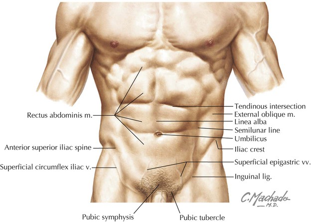
The rectus abdominis is positioned between the ribs and the pubic bone at the front of the pelvis, and is actually made up of 8 distinct muscle bellies.
Abdominal muscle anatomy male i mean, the abs are the muscle. The abdomen extends as far superiorly as the seventh costal cartilage. This diagram depicts male abdomen anatomy. The large intestine connects to the small intestine in the lower right section of the abdominal cavity. Human anatomy diagrams show internal organs, cells, systems, conditions, symptoms and sickness information and/or tips for healthy living. One of the easiest ways to tell if your pain is caused by a hernia or pulled stomach muscle is if you have a bulge or not. Ribs, sternum, infrasternal angle and costal margin: Muscles are the only tissue in the body that has the ability to contract and therefore move the other parts of the body. The rectus abdominis is positioned between the ribs and the pubic bone at the front of the pelvis, and is actually made up of 8 distinct muscle bellies. Muscles often contract to hold the body still or in a particular position rather than to cause. Working as a team, these muscles contract to flex, laterally bend, and rotate the torso. The maintenance of posture and body position. Inflammation of the covering of the abdominal structures, causing abdominal wall rigidity and severe pain.
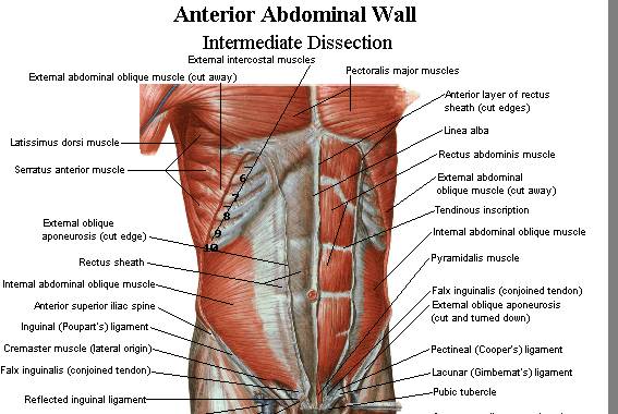
One of the easiest ways to tell if your pain is caused by a hernia or pulled stomach muscle is if you have a bulge or not. They collectively form part of the boundaries of the abdominal cavity. The rectus abdominis muscle, also known as the abdominal muscle, is a paired muscle running vertically on each side of the anterior wall of the human abdomen. Anatomy of lower limb arteries 12 photos of the anatomy of lower limb arteries cross sectional anatomy of lower limb arteries, ct anatomy of lower limb arteries, lower limb arterial anatomy images, lower limb arteries instant anatomy, teach me anatomy arteries of lower limb, human anatomy, cross sectional. The external obliques, the internal obliques, the transversus abdominis, and the rectus abdominis (figure 1, figure 2, and table 1).
60 Top Full Stomach Stock Illustrations, Clip art ... from media.gettyimages.com The groin is the area in the body where the upper thighs meet the lowest part of the abdomen. The fourth layer in the midregion is the rectus abdominis, which has vertically running muscle fibres that flex the trunk and stabilize the pelvis. They collectively form part of the boundaries of the abdominal cavity. A muscle strain occurs when the muscle is stretched too far. The images are labeled, providing an invaluable medical tool. Possible causes of lower abdominal pain in men 1. Here is a diagram that shows where each one is located anatomy of the abdominal cavity: These muscles of the anterolateral abdominal wall can be divided into four groups:
See abdominal muscle stock video clips.
The external oblique, internal oblique, transverse abdominal, and cremaster muscles form the lateral abdominal muscle group. The male abdomen below the belly button contains part of the stomach, colon, appendix and rectum. Anatomy of lower limb arteries 12 photos of the anatomy of lower limb arteries cross sectional anatomy of lower limb arteries, ct anatomy of lower limb arteries, lower limb arterial anatomy images, lower limb arteries instant anatomy, teach me anatomy arteries of lower limb, human anatomy, cross sectional. Skin, superficial fascia, muscles and associated fascia, and parietal peritoneum. The abdomen extends as far superiorly as the seventh costal cartilage. The muscle strain is accompanied by increased pressure surrounding them and may make the abdominal pain worse. When this occurs the muscle fibers are torn. Extending across the anterior surface of the body from the superior border of the pelvis to the inferior border of the ribcage are the muscles of the abdominal wall, including the transverse and rectus abdominis and the internal and external obliques. We're going to take apart a plastic anatomy model and see what we can find in the abdomen. In the rear of the abdomen are the back muscles and spine. The anterolateral abdominal wall consists of four main layers (external to internal): Occasionally, in severe injuries, the muscle. Anatomy of the abdominal cavity and the male pelvis:
The anterolateral abdominal wall consists of four main layers (external to internal): This tool provides access to a ct atlas in the axial plane, allowing the user to interactively learn abdominal anatomy. Human anatomy diagrams show internal organs, cells, systems, conditions, symptoms and sickness information and/or tips for healthy living. No need to register, buy now! Ribs, sternum, infrasternal angle and costal margin:
Abdomen | Basicmedical Key from basicmedicalkey.com These muscles of the anterolateral abdominal wall can be divided into four groups: The rectus abdominis is positioned between the ribs and the pubic bone at the front of the pelvis, and is actually made up of 8 distinct muscle bellies. We're going to take apart a plastic anatomy model and see what we can find in the abdomen. How to view anatomical labels. Anterolateral abdominal wall muscles external obliques muscle, the most superficial anterolateral abdominal muscle its fibers run inferomedially, unilateral action results in ipsilateral side flexion and contralateral rotation of the trunk bilateral action to flex the vertebral column by drawing the pubis towards the xiphoid proces s. Working as a team, these muscles contract to flex, laterally bend, and rotate the torso. The abdominal muscles play critical roles in spinal stability, breathing, protection of your internal organs and movements of your core. The anterolateral abdominal wall consists of four main layers (external to internal):
The anterior abdominal muscles are part of the musculature that contributes to the anterolateral abdominal wall, along with the lateral abdominal muscles on either side.
The pain is caused by increased strain that abdominal muscles are under in constipation. Anatomy of male abdomen, find out more about anatomy of male abdomen. Find the perfect male abdomen anatomy stock photo. Abdominal muscle anatomy male i mean, the abs are the muscle. Skin, superficial fascia, muscles and associated fascia, and parietal peritoneum. The rectus abdominis muscle, also known as the abdominal muscle, is a paired muscle running vertically on each side of the anterior wall of the human abdomen. Identify the major skeletal landmarks of the abdominopelvic cavity. They collectively form part of the boundaries of the abdominal cavity. Possible causes of lower abdominal pain in men 1. The muscles of the anterior abdominal wall are located near the midline between the costal margin superiorly and the pubis inferiorly. This tool provides access to a ct atlas in the axial plane, allowing the user to interactively learn abdominal anatomy. The images are labeled, providing an invaluable medical tool. See abdominal muscle stock video clips.